Home

coadă zenit Îndepărta right marker us probe uite dialect îngust

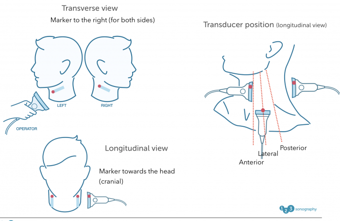
4. B mode imaging – Carotid artery | 123 Sonography

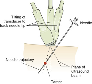
How To: Ultrasound Guidance for Central Venous Access Part 2 Case Study  Video - YouTube
How To: Ultrasound Guidance for Central Venous Access Part 2 Case Study Video - YouTube

Ford Probe 1989 Right Side Marker Lamp w/ Turbo Amber NOS OEM Vintage | eBay

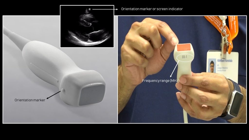
Orientation | Radiology Key

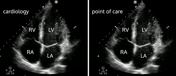
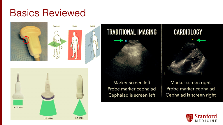
Stanford Peds Chiefs on X: "2/ Basics reviewed: Remember your different  kind of probes. Note the difference in side where the probe marker is for  echo's compared to traditional US imaging. Also
Stanford Peds Chiefs on X: "2/ Basics reviewed: Remember your different kind of probes. Note the difference in side where the probe marker is for echo's compared to traditional US imaging. Also

BASIC CHARACTERISTICS OF THE ULTRASOUND EQUIPMENT

A Novel Ultrasound Probe Spatial Calibration Method Using a Combined  Phantom and Stylus - Ultrasound in Medicine and Biology
A Novel Ultrasound Probe Spatial Calibration Method Using a Combined Phantom and Stylus - Ultrasound in Medicine and Biology
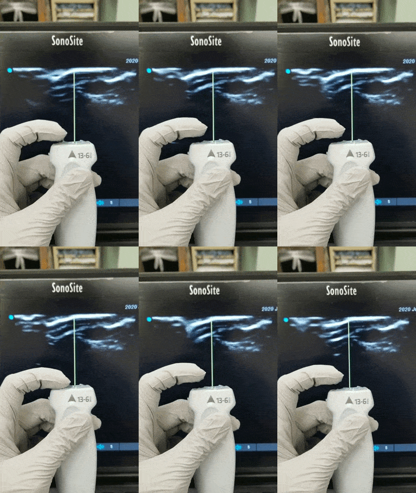
Basic probe positioning and screen orientation for an arterial du |  Medmastery
Basic probe positioning and screen orientation for an arterial du | Medmastery

UOTW #33 Answer - Core Ultrasound

Image Orientation and Transducer Position - YouTube

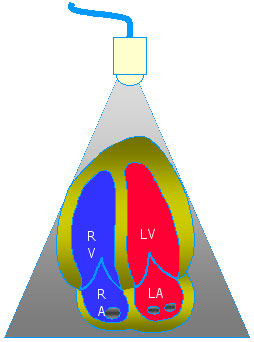
Cardiac ultrasound - WikEM

Ultrasound probe with markers and optical tracker | Download Scientific  Diagram
Ultrasound probe with markers and optical tracker | Download Scientific Diagram

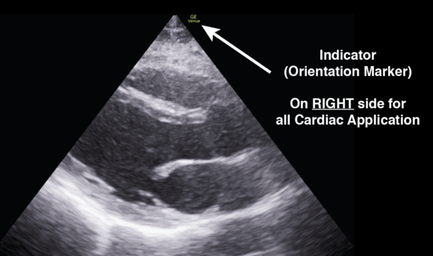
POCUS 101 on X: "(9) Understand how the Indicator (orientation marker) on  the probe correlates with the ultrasound image. Standard: Left side of the  screen Cardiac: Right side of the screen 👉🔗https://t.co/pt5YwCmqlt
POCUS 101 on X: "(9) Understand how the Indicator (orientation marker) on the probe correlates with the ultrasound image. Standard: Left side of the screen Cardiac: Right side of the screen 👉🔗https://t.co/pt5YwCmqlt

Ultrasound Teaching Cases

Probe marker should be in the same direction as the marker on the... |  Download Scientific Diagram
Probe marker should be in the same direction as the marker on the... | Download Scientific Diagram

Orientation | Radiology Key

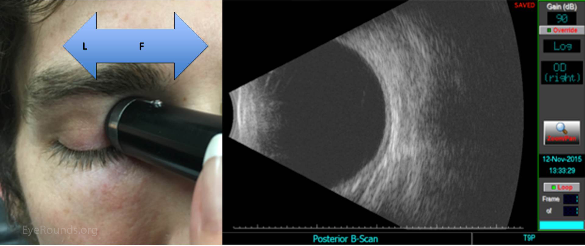
Ocular Ultrasound: A Quick Reference Guide for the On-Call Physician

Basic probe positioning and screen orientation for an arterial du |  Medmastery
Basic probe positioning and screen orientation for an arterial du | Medmastery

EMERGENCY ULTRASOUND | Anesthesia Key

Introduction to Focused Cardiac Ultrasound: The Parasternal Long Axis View  - Renal Fellow Network
Introduction to Focused Cardiac Ultrasound: The Parasternal Long Axis View - Renal Fellow Network

Probe Orientation - RCEMLearning India

Basic probe positioning and screen orientation for an arterial du |  Medmastery
Basic probe positioning and screen orientation for an arterial du | Medmastery

Probe marker should be in the same direction as the marker on the... |  Download Scientific Diagram
Probe marker should be in the same direction as the marker on the... | Download Scientific Diagram

Orientation | Radiology Key

Point-of-care ultrasound - Knowledge @ AMBOSS

Ultrasound-Guided Peripheral IV Insertion, Placement, and Access Made Easy  - POCUS 101
Ultrasound-Guided Peripheral IV Insertion, Placement, and Access Made Easy - POCUS 101

PROBECRAFT — MMHEME
PROBECRAFT — MMHEME

Scanning School - Cardiac — Taming the SRU